
 Image
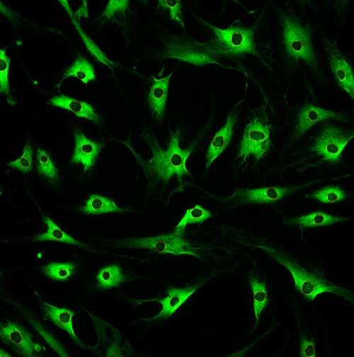 Single cells
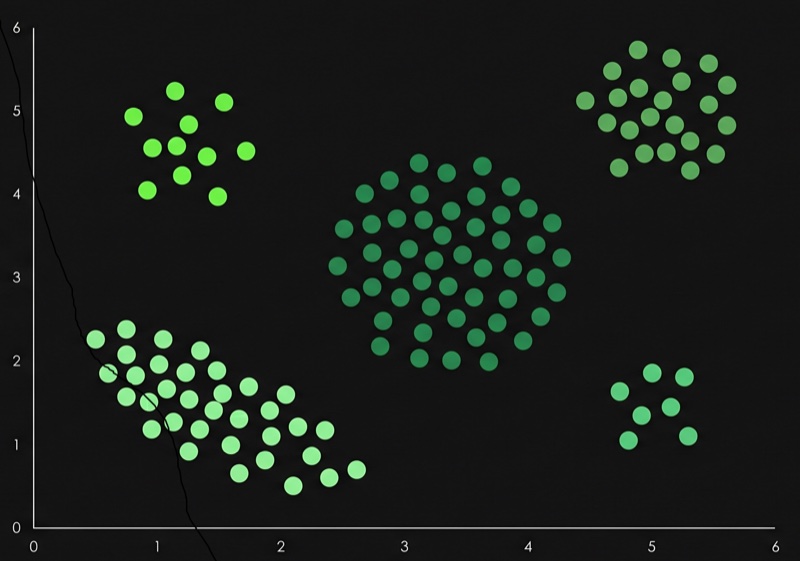 Cluster
 Profile
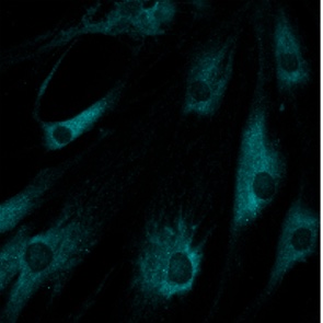
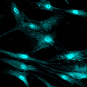
Image evidence · model description
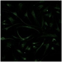
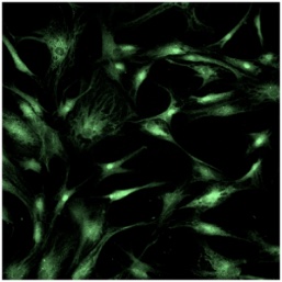
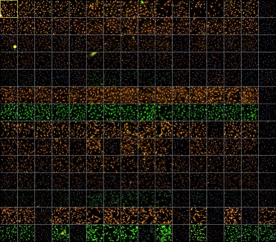
Test cells show a dramatic shift in staining patterns compared to reference cells. The most striking change is a massive increase in cells displaying moderate diffuse nuclear staining with rounded dark nuclear holes accompanied by moderate granular cytoplasmic staining, indicating the emergence of a phenotype virtually absent in the reference group. Concurrently, there is a near-complete loss of cells exhibiting weak diffuse nuclear staining paired with moderate diffuse cytoplasmic staining, a phenotype that predominates in the reference population.
Biological meaning · interpretation
Non-phosphorylated Src translocated in RA-FLS cells from the cytoplasm to the nucleus. Nuclear, inactive, Src scaffolds the splicing machinery, converging with the alternative splicing hypothesis implied by SRPK1-SRSF activation.
Experimental contextSrc · primary FLS · healthy donor (reference) vs RA patient (test)